A 78-year-old woman falls at home and sustains a displaced subcapital fracture of the femoral neck. The surgeon chooses hemiarthroplasty rather than internal fixation.
Damage to which vessel best explains this decision?
How sure are you?
Revise by topic
Lower limb anatomy runs from the gluteal region and hip joint, through the femoral triangle and adductor canal, to the popliteal fossa, the four compartments of the leg, the ankle and the foot, with the arterial tree, the venous system and the lymphatics of the limb alongside. Applied surgical anatomy is 75 of the 180 Paper 1 questions; the lower limb contributes 7 of them. The domain is built on relations that matter at operation and after injury: what lies in the femoral triangle and in what order, where the sciatic nerve runs in relation to the hip, where the common fibular nerve is exposed at the neck of the fibula, which nerve and artery supply each compartment, and how the great saphenous vein relates to the medial malleolus and the saphenofemoral junction. Questions typically present a fracture, a dislocation, a vascular procedure or a varicose vein operation and ask which structure explains the finding or is at risk.
Topics in this domain
From the free sample — original, referenced, with the full explanation. Rate your confidence first.
A 78-year-old woman falls at home and sustains a displaced subcapital fracture of the femoral neck. The surgeon chooses hemiarthroplasty rather than internal fixation.
Damage to which vessel best explains this decision?
Drag to rotate, tap the hotspots, press play.
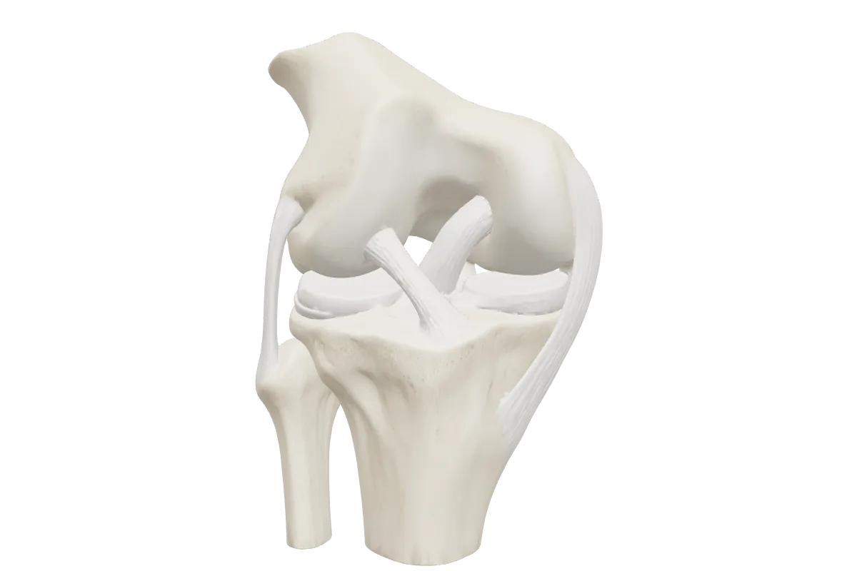
Also in this domain: Blood supply of the femoral head, Sciatic nerve & short external rotators, Ankle ligaments & syndesmosis, Foot skeleton, Femoral triangle & canal (coming soon), Popliteal fossa (coming soon)
From lateral to medial, the femoral nerve, the femoral artery, the femoral vein and the femoral canal with its lymphatics and the node of Cloquet. The triangle is bounded by the inguinal ligament, the medial border of sartorius and the medial border of adductor longus, with a floor of iliopsoas, pectineus and adductor longus.
From superficial to deep: the tibial nerve, the popliteal vein and the popliteal artery, which lies deepest against the femur and the capsule of the knee. That is why the artery is injured in posterior knee dislocations and supracondylar femoral fractures. The common fibular nerve runs along the medial border of biceps femoris at the lateral edge of the fossa.
The common fibular (peroneal) nerve. Injury gives foot drop from paralysis of the anterior and lateral compartments — loss of dorsiflexion and eversion — with a high-stepping gait and sensory loss over the dorsum of the foot and the lateral leg.
The ICBSE blueprint lists 7 of the 180 Paper 1 questions under the lower limb, within the 75 for applied surgical anatomy. The same anatomy underpins Paper 2 questions on hip fracture, compartment syndrome, varicose veins and limb ischaemia.
Create a free account, filter the bank to this domain and rate your confidence as you go. Upgrade when you want the full bank and two-paper mocks.