Skip to content

Revise by topic

Spine & embryology for MRCS Part A

This domain gathers three blueprint lines that sit outside regional anatomy. Spine and spinal cord covers the vertebrae, discs and ligaments, the cord with its roots, dermatomes and myotomes, and the levels at which the cord and the dural sac end. Surgically related embryology covers the rotation of the gut and the malrotations and atresias that follow, the pharyngeal arches and pouches and the descent of the thyroid, the heart and great vessels, the urogenital system, and the limbs, body wall and diaphragm. Surface and imaging anatomy covers the landmarks and vertebral levels a surgeon uses to plan an incision or read a radiograph, and the cross-sectional anatomy of CT and MRI. Applied surgical anatomy is 75 of the 180 Paper 1 questions; these three lines contribute 15 of them — spine 3, embryology 8 and surface and imaging anatomy 4. Embryology is tested through the congenital conditions it explains rather than for its own sake.

150 questions in the bank · 3 topics Updated 6 October 2026

Topics in this domain

  • Spine & spinal cord30
  • Surgically related embryology80
  • Surface & imaging anatomy40

What the paper tends to test

  • Questions typically test spinal levels: where the cord ends in the adult and the neonate, where a lumbar puncture is safe, and which layers the needle passes through.
  • Expect dermatomes, myotomes and reflexes to be used to localise a root lesion — the L4, L5 and S1 patterns of a lumbar disc prolapse and the C5–T1 patterns in the arm.
  • Embryology is asked through malformations: malrotation and volvulus, Meckel’s diverticulum, tracheo-oesophageal fistula, congenital diaphragmatic hernia, and the structures derived from each pharyngeal arch and pouch.
  • Thyroid descent questions test the thyroglossal duct and cyst, a lingual thyroid and the pyramidal lobe; parathyroid questions test why the inferior glands are the variable ones.
  • Surface anatomy questions tend to ask for the vertebral level of a landmark — the transpyloric plane, the sternal angle, the aortic bifurcation — or the surface marking of an organ.
  • Imaging anatomy is examined as a described CT, MRI or plain film with a structure to identify or a level to name.
  • Cardiac embryology is tested through the fetal circulation, the shunts that close at birth and the common septal defects.

Classic traps and discriminators

  • The spinal cord ends at about L1–L2 in the adult (L3 in the neonate) and the dural sac at S2, so a lumbar puncture at L3–L4 or L4–L5 enters the subarachnoid space among the roots of the cauda equina without reaching the cord.
  • The transpyloric plane (L1) crosses the pylorus, the duodenojejunal flexure, the neck of the pancreas, the fundus of the gallbladder, the hila of the kidneys and the origin of the superior mesenteric artery; the aorta bifurcates at L4 and the inferior vena cava forms at L5.
  • The midgut rotates 270° counter-clockwise around the superior mesenteric artery; incomplete rotation leaves a narrow mesenteric base and Ladd’s bands, so bilious vomiting in a neonate is malrotation with volvulus until proved otherwise.
  • The thyroid descends from the foramen caecum along the thyroglossal duct, which is why a thyroglossal cyst is midline and rises on protrusion of the tongue; the inferior parathyroids come from the third pouch with the thymus and the superior from the fourth, so the inferior glands vary more in position.
  • A Meckel’s diverticulum is the remnant of the vitellointestinal duct: on the antimesenteric border of the ileum, about 60 cm (2 feet) from the ileocaecal valve, in about 2% of people, and often lined by ectopic gastric or pancreatic mucosa that bleeds or perforates.
  • The sternal angle (of Louis) marks the second costal cartilage and the T4–T5 disc: the beginning and end of the aortic arch, the bifurcation of the trachea, the boundary between the superior and inferior mediastinum and the point where the azygos vein enters the superior vena cava.

Free spine & embryology practice question

From the free sample — original, referenced, with the full explanation. Rate your confidence first.

medium

A 24-year-old man dives into shallow water and strikes the crown of his head. CT shows fractures of both the anterior and posterior arches of the atlas, with the lateral masses of C1 displaced laterally by a combined total of 8 mm on the open-mouth view.

Which ligament is most likely to have been disrupted, making this injury unstable?

How sure are you?
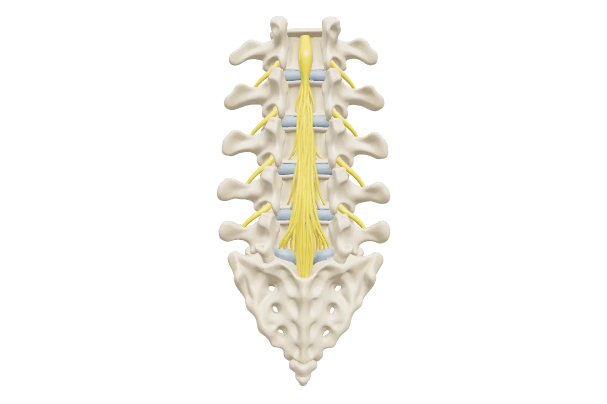
Interactive 3D: Lumbar spine & cauda equina

Drag to rotate, tap the hotspots, press play.

Lumbar spine & cauda equina — still render

Also in this domain: Lumbar vertebra, Atlas & axis (C1–C2), Lumbar vertebra & spinal cord (coming soon)

Spine & embryology questions, answered

At what level does the spinal cord end?

At the lower border of L1 or the upper border of L2 in most adults, and about L3 in the neonate. The subarachnoid space and dural sac continue to S2, so the cauda equina, not the cord, occupies the space below L2 — the basis of a safe lumbar puncture or spinal anaesthetic.

Which structures come from each pharyngeal arch?

First arch (mandibular nerve, V3): maxilla, mandible, malleus, incus and the muscles of mastication. Second (facial nerve): stapes, styloid process, lesser horn of the hyoid and the muscles of facial expression. Third (glossopharyngeal): greater horn and body of the hyoid, stylopharyngeus. Fourth and sixth (vagus): the laryngeal cartilages and the pharyngeal and laryngeal muscles, with the superior laryngeal nerve from the fourth and the recurrent laryngeal nerve from the sixth.

Why is a Meckel’s diverticulum important?

Because it mimics appendicitis, bleeds from ectopic gastric mucosa, acts as a lead point for intussusception or a band for obstruction, and may be found incidentally at laparotomy. The ‘rule of twos’ — 2% of the population, about 2 feet from the ileocaecal valve, 2 inches long, often symptomatic before the age of 2 — is the usual exam framing.

How many questions do spine, embryology and surface anatomy contribute?

The ICBSE blueprint gives these three lines 15 of the 180 Paper 1 questions: spine 3, surgically related embryology 8, and surface and imaging anatomy 4, within the 75 for applied surgical anatomy.

Drill spine & embryology tonight

Create a free account, filter the bank to this domain and rate your confidence as you go. Upgrade when you want the full bank and two-paper mocks.