A 78-year-old woman falls at home and sustains a displaced subcapital fracture of the femoral neck. The surgeon chooses hemiarthroplasty rather than internal fixation.
Damage to which vessel best explains this decision?
How sure are you?
Lower limb · Free article
Femoral triangle for MRCS Part A: boundaries, roof and floor, contents in order, the femoral sheath and canal, femoral hernia and profunda branches.
| Boundary | Structure |
|---|---|
| Superior (base) | Inguinal ligament |
| Lateral | Medial border of sartorius |
| Medial | Medial border of adductor longus |
| Apex | Where sartorius crosses adductor longus; continuous with the adductor canal |
| Roof | Skin, superficial fascia (containing the superficial inguinal nodes, the great saphenous vein and the superficial branches of the femoral artery) and fascia lata, with the cribriform fascia over the saphenous opening |
| Floor (lateral to medial) | Iliopsoas, pectineus and adductor longus |
The floor is a gutter: iliopsoas and pectineus slope towards each other, so the femoral vessels lie in a groove between them. The great saphenous vein pierces the cribriform fascia at the saphenous opening, a few centimetres below and lateral to the pubic tubercle, to join the femoral vein at the saphenofemoral junction.
From lateral to medial the contents are the femoral nerve, the femoral artery, the femoral vein and the femoral canal (often remembered as NAVY, with the Y for the empty space). The femoral sheath is a funnel-shaped extension of the abdominal fascia: transversalis fascia in front and iliac fascia behind. It extends about 3–4 cm below the inguinal ligament and has three compartments.
| Landmark | Definition | What lies there |
|---|---|---|
| Mid-inguinal point | Halfway between the anterior superior iliac spine and the pubic symphysis | The femoral artery (the femoral pulse) |
| Midpoint of the inguinal ligament | Halfway between the anterior superior iliac spine and the pubic tubercle | The deep inguinal ring, about 1–2 cm above it |
For vascular access, the femoral vein lies medial to the pulse and the femoral nerve lateral to it. Puncturing the artery over the femoral head lets it be compressed afterwards: too high risks retroperitoneal haemorrhage, and too low risks the bifurcation, a pseudoaneurysm or an arteriovenous fistula.
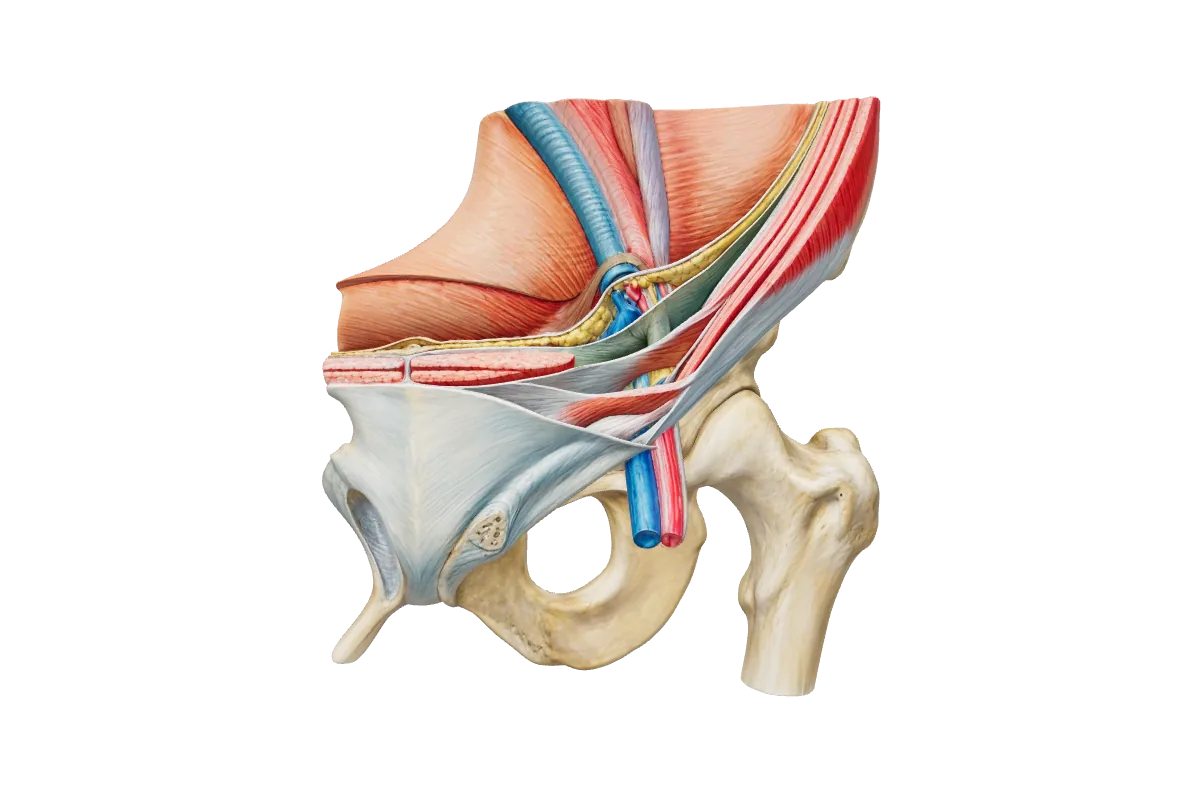
The femoral canal is the medial compartment of the sheath, about 1–2 cm long. Its upper opening, the femoral ring, has four boundaries:
| Side | Boundary |
|---|---|
| Anterior | Inguinal ligament |
| Posterior | Pectineal ligament (Cooper’s ligament) over the superior pubic ramus |
| Medial | Lacunar ligament (Gimbernat’s ligament) |
| Lateral | Femoral vein |
Three of the four boundaries are unyielding, so a femoral hernia has a narrow neck and a high risk of incarceration, strangulation and Richter’s hernia, in which only part of the bowel wall is trapped. The hernia passes down the canal, emerges through the saphenous opening and may then turn upwards over the inguinal ligament. Femoral hernias are commoner in women than in men, but even in women inguinal hernias are more common than femoral ones.
The adductor (subsartorial, or Hunter’s) canal runs from the apex of the femoral triangle to the adductor hiatus in adductor magnus. Vastus medialis lies anterolaterally, adductor longus and adductor magnus posteriorly, and sartorius over the roof. It contains the femoral artery and vein, the saphenous nerve and the nerve to vastus medialis. At the hiatus the femoral vessels become the popliteal vessels. An adductor canal block anaesthetises the saphenous nerve for knee surgery while sparing most of quadriceps.
Test yourself
A 78-year-old woman falls at home and sustains a displaced subcapital fracture of the femoral neck. The surgeon chooses hemiarthroplasty rather than internal fixation.
Damage to which vessel best explains this decision?
Test yourself
A 72-year-old woman taking apixaban develops sudden lower abdominal pain after a bout of coughing. There is a tender, non-pulsatile mass in the right lower quadrant that remains palpable and becomes more tender when she lifts her head from the pillow.
Which vessel is the most likely source of bleeding?
The inguinal ligament superiorly, the medial border of sartorius laterally and the medial border of adductor longus medially. The roof is fascia lata with the cribriform fascia; the floor is iliopsoas, pectineus and adductor longus.
No. The sheath contains the femoral artery, the femoral vein and the femoral canal. The femoral nerve lies lateral to the sheath, in the groove between iliacus and psoas.
The inguinal ligament anteriorly, the pectineal (Cooper’s) ligament posteriorly, the lacunar ligament medially and the femoral vein laterally.
By its relation to the pubic tubercle. A femoral hernia lies below and lateral to the tubercle; an inguinal hernia lies above and medial to it.
At the mid-inguinal point, halfway between the anterior superior iliac spine and the pubic symphysis. The midpoint of the inguinal ligament, halfway between the anterior superior iliac spine and the pubic tubercle, marks the deep inguinal ring.
Written for revision in the MRCS Part A style under our editorial standards. It is not clinical advice: always confirm against current guidance. Spotted an error? Tell us.
Abdomen & pelvis
Inguinal canal anatomy for MRCS Part A: walls, rings, cord coverings, indirect versus direct hernias, the femoral canal, surface landmarks and nerves at risk.
ReadPathology
Nerve injury for MRCS Part A: nerve structure, Seddon and Sunderland grades, Wallerian degeneration, regeneration, nerve conduction timing and repair.
ReadAbdomen & pelvis
Calot's triangle for MRCS Part A: hepatocystic boundaries and contents, cystic artery and duct variants, the critical view of safety and bile duct injury.
ReadDrill lower limb with curriculum-mapped questions, rate your confidence, and watch the gap close on your dashboard.