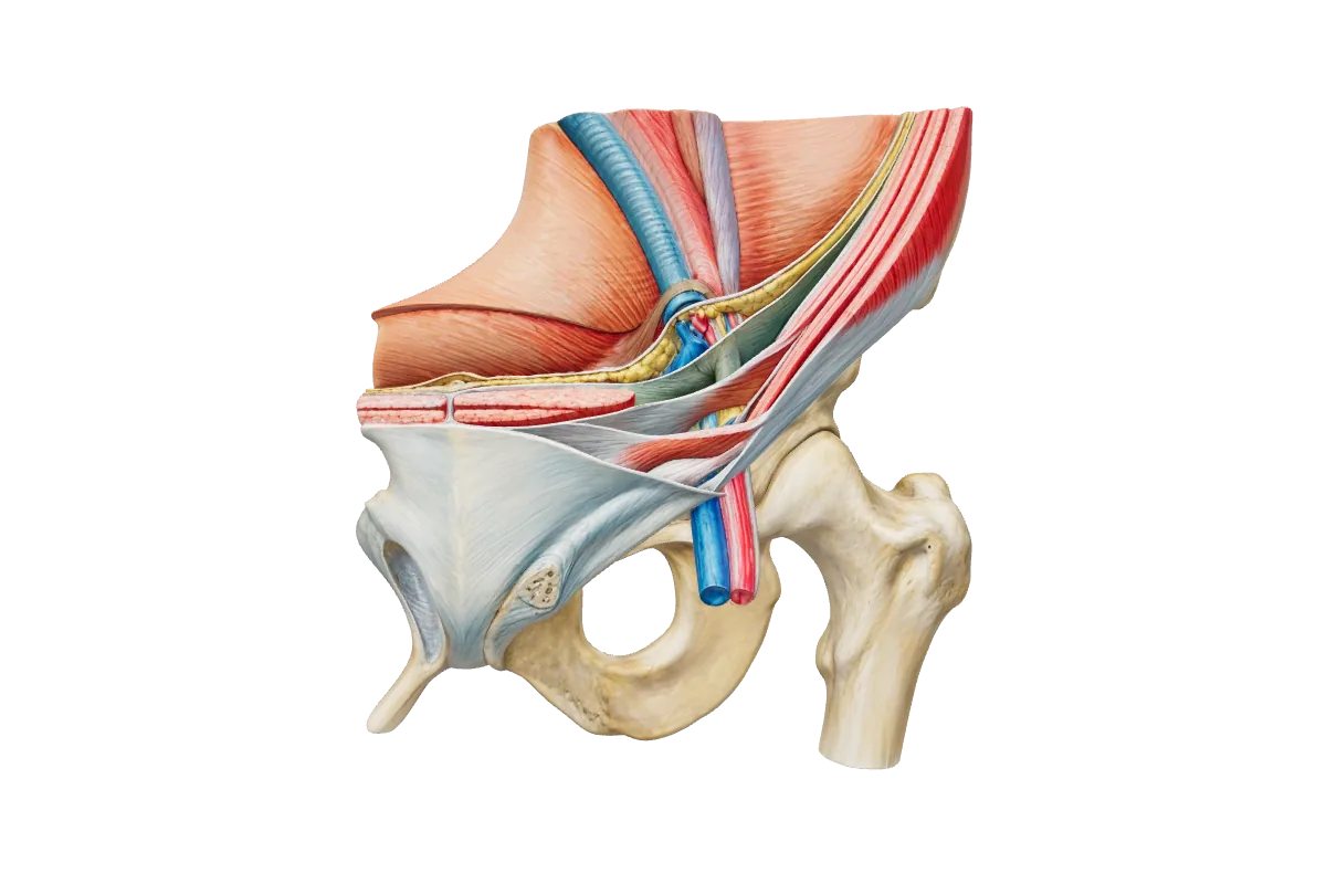
Abdomen, pelvis and perineum is the largest anatomical domain. Applied surgical anatomy is 75 of the 180 Paper 1 questions, and this domain contributes 21 of them: abdominal wall and peritoneal cavity 6, abdominal viscera 6, abdominal vessels 3, pelvis 4 and perineum 2. It covers the layers of the abdominal wall and the inguinal canal, the peritoneal reflections, mesenteries and the lesser sac, the foregut, midgut and hindgut organs with their blood supply, the liver, biliary tree and Calot’s triangle, the pancreas and spleen, the kidneys, ureters and adrenals, the aorta and its branches, the portal system and its portosystemic anastomoses, the bony pelvis and pelvic floor, the rectum, bladder, prostate and uterus with their vessels and nerves, and the anal canal and urogenital triangle. Questions are rooted in operations — hernia repair, cholecystectomy, colectomy, nephrectomy, anterior resection — and in the relations a surgeon has to know to avoid the ureter, the common bile duct or the pelvic nerves.
210 questions in the bank · 5 topics Updated 6 October 2026
Questions typically test the inguinal canal and hernia anatomy: the walls of the canal, the position of the deep ring, and what separates a direct from an indirect and a femoral hernia.
Expect Calot’s triangle — its boundaries, the cystic artery and the common variations — framed as the anatomy of a safe laparoscopic cholecystectomy.
Blood supply questions follow the embryological divisions: the coeliac, superior mesenteric and inferior mesenteric territories, the marginal artery and the watershed areas.
The ureter is examined by its course and its relations to the gonadal vessels, the iliac vessels and the uterine artery, and by where it is at risk in colorectal, gynaecological and vascular surgery.
Pelvic questions tend to test the pelvic floor, the autonomic plexuses and the structures damaged during rectal mobilisation or a radical prostatectomy.
Anal canal questions turn on the dentate line — the change in epithelium, blood supply, venous and lymphatic drainage and nerve supply above and below it.
Portal hypertension is examined through the sites of portosystemic anastomosis and what each produces.
Classic traps and discriminators
The deep inguinal ring lies lateral to the inferior epigastric vessels, so an indirect hernia emerges lateral to them and a direct hernia medial to them through Hesselbach’s triangle. A femoral hernia lies below and lateral to the pubic tubercle; an inguinal hernia above and medial to it.
Calot’s triangle is bounded by the cystic duct, the common hepatic duct and the inferior surface of the liver; the cystic artery usually arises from the right hepatic artery within it, and the critical view of safety must be obtained before anything is clipped or divided.
The ureter crosses the pelvic brim at the bifurcation of the common iliac artery and passes beneath the uterine artery (‘water under the bridge’). It is at risk at the pelvic brim, at the ligation of the inferior mesenteric artery and the ovarian pedicle, and at the uterine artery close to the lateral fornix.
Above the dentate line the anal canal has columnar epithelium, is supplied by the superior rectal artery, drains to the portal system and the internal iliac nodes and is insensitive to pain; below it the epithelium is squamous, the venous drainage is systemic, the lymph goes to the superficial inguinal nodes and the inferior rectal (pudendal) nerve makes it exquisitely sensitive.
Portosystemic anastomoses: left gastric with the oesophageal veins (varices), superior rectal with the middle and inferior rectal veins (rectal varices, which are not haemorrhoids), paraumbilical with the epigastric veins (caput medusae) and the retroperitoneal veins of Retzius.
The tail of the pancreas lies in the lienorenal (splenorenal) ligament with the splenic vessels, so it is at risk during splenectomy; the spleen’s other relations are the stomach, the left kidney and the splenic flexure, and its hilum faces the lesser sac.
Free abdomen & pelvis practice questions
From the free sample — original, referenced, with the full explanation. Rate your confidence first.
A 72-year-old woman taking apixaban develops sudden lower abdominal pain after a bout of coughing. There is a tender, non-pulsatile mass in the right lower quadrant that remains palpable and becomes more tender when she lifts her head from the pillow.
Which vessel is the most likely source of bleeding?
How sure are you?
Explanation
A mass that persists and becomes more tender on tensing the abdominal wall (positive Fothergill's and Carnett's signs) in an anticoagulated patient is a rectus sheath haematoma. Most arise below the arcuate line, where branches of the inferior epigastric artery enter the sheath on the deep surface of the muscle and where, lacking a posterior sheath, bleeding can spread widely into the extraperitoneal and prevesical tissues. The superior epigastric artery supplies the upper rectus above the umbilicus, where haematomas are small and confined between tendinous intersections. The deep circumflex iliac artery runs along the iliac crest in the lateral wall, and the superficial epigastric artery lies in the subcutaneous fat. External iliac bleeding would produce a retroperitoneal haematoma with haemodynamic collapse rather than a wall mass. Reference: Bailey & Love 28e; Last's Anatomy 12e.
A 40-year-old man has painless bright-red bleeding after defaecation. Proctoscopy shows prolapsing haemorrhoids arising above the dentate line.
Into which vein do the dilated vessels principally drain?
How sure are you?
Explanation
Internal haemorrhoids are enlargements of the anal cushions, vascular plexuses in the submucosa above the dentate line at the left lateral, right anterior and right posterior positions (3, 7 and 11 o'clock in the lithotomy position). They are fed by terminal branches of the superior rectal artery and drain through the superior rectal vein to the inferior mesenteric vein and so to the portal system, which is why the anal canal is a site of portosystemic anastomosis. The middle rectal veins drain the muscular wall of the lower rectum to the internal iliac veins, and the inferior rectal veins drain the canal below the dentate line to the internal pudendal veins, both systemic. The median sacral vein drains the back of the rectum and sacrum. Reference: Last's Anatomy 12e.
Anterior: the external oblique aponeurosis, reinforced laterally by internal oblique. Posterior: transversalis fascia, reinforced medially by the conjoint tendon. Roof: the arching fibres of internal oblique and transversus abdominis. Floor: the inguinal ligament, with the lacunar ligament medially. It carries the spermatic cord or the round ligament, and the ilioinguinal nerve.
How do I tell a direct from an indirect inguinal hernia?+
By its relation to the inferior epigastric vessels at operation: an indirect hernia enters the deep ring lateral to them and lies within the coverings of the cord; a direct hernia bulges through the posterior wall medial to them, in Hesselbach’s triangle. Clinical tests such as occluding the deep ring are unreliable, which is a favourite point for a question.
Where is the ureter at risk in surgery?+
At the pelvic brim where it crosses the common iliac bifurcation, during ligation of the inferior mesenteric artery or the ovarian pedicle, where it passes under the uterine artery near the lateral vaginal fornix, and at the ureterovesical junction. It is identified by its peristalsis and its position on the medial leaf of the mesentery.
How many abdomen, pelvis and perineum questions are in MRCS Part A?+
The ICBSE blueprint gives this domain 21 of the 180 Paper 1 questions: abdominal wall and cavity 6, viscera 6, vessels 3, pelvis 4 and perineum 2 — the largest anatomical domain. Hernias, the acute abdomen and colorectal disease are examined again, as clinical conditions, in Paper 2.
Drill abdomen & pelvis tonight
Create a free account, filter the bank to this domain and rate your confidence as you go. Upgrade when you want the full bank and two-paper mocks.