Inguinal canal and hernia anatomy: walls, rings, the cord and the femoral canal
Inguinal canal anatomy for MRCS Part A: walls, rings, cord coverings, indirect versus direct hernias, the femoral canal, surface landmarks and nerves at risk.
An oblique passage through the lower abdominal wall
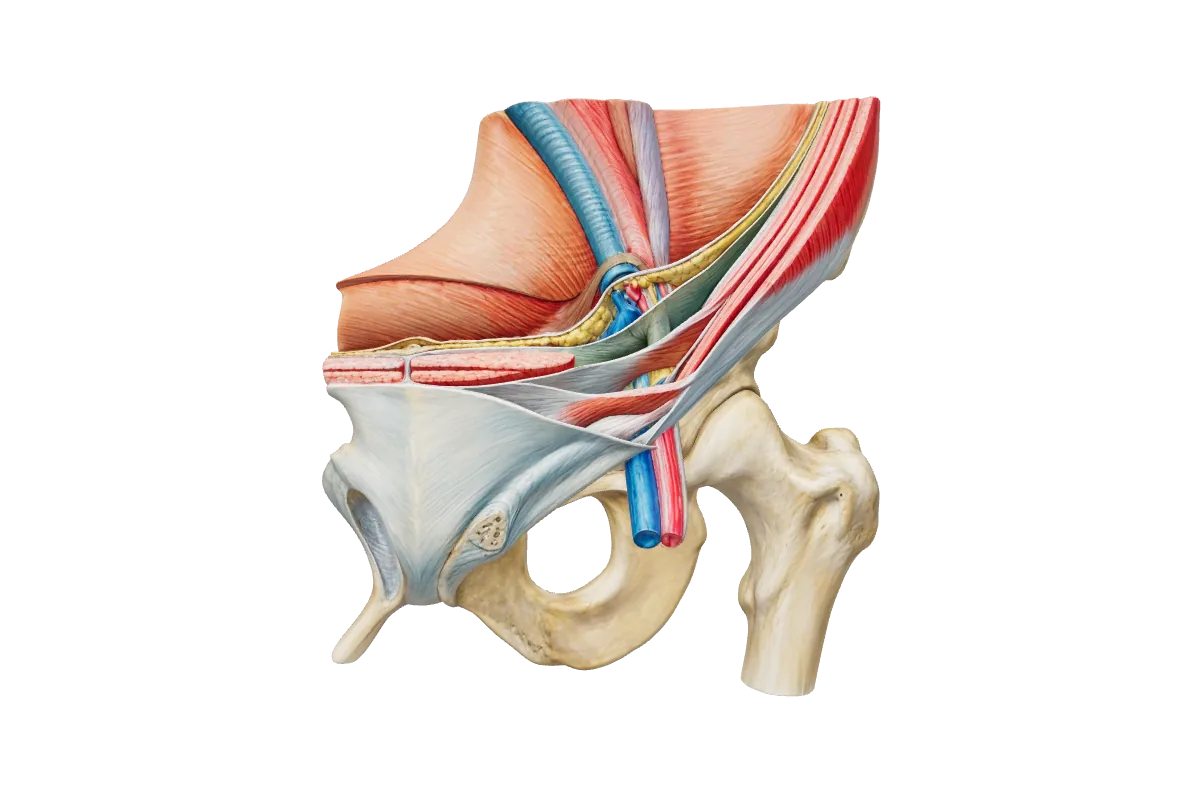
The inguinal canal is an oblique passage about 4 cm long through the lower anterior abdominal wall, immediately above the medial half of the inguinal ligament. It exists because the testis, or the round ligament, passed through the wall during development and dragged a sleeve of each layer with it. Its entrance is the deep (internal) ring, an oval opening in the transversalis fascia about 1.25 cm above the midpoint of the inguinal ligament and immediately lateral to the inferior epigastric vessels. Its exit is the superficial (external) ring, a triangular split in the external oblique aponeurosis resting on the pubic crest, just above and medial to the pubic tubercle, to which its lateral crus is attached. Because the rings are not opposite one another, a rise in intra-abdominal pressure flattens the canal rather than opening it: the arching fibres of internal oblique and transversus drop like a shutter across the deep ring, and the conjoint tendon braces the wall behind the superficial ring. The canal transmits the spermatic cord or the round ligament and, in both sexes, the ilioinguinal nerve, which enters not through the deep ring but by piercing internal oblique part-way along, and leaves through the superficial ring lying on the front of the cord.
Interactive 3D · Inguinal canal & hernia anatomy. The left groin from the front: find the deep ring, then the inguinal ligament with the femoral artery, vein and canal beneath it.Open full screen
Each wall is a different layer of the abdominal wall; the cord and an indirect sac carry coverings derived from them.
Wall
Formed by
What it means
Anterior
External oblique aponeurosis throughout; the lowest fibres of internal oblique in front of the lateral third, where they arise from the inguinal ligament
Reinforces the deep ring, the lateral weak point
Posterior
Transversalis fascia throughout; the conjoint tendon (falx inguinalis) behind the medial third
Reinforces the superficial ring; the unsupported middle is Hesselbach's triangle, where a direct hernia appears
Roof
Arching lower fibres of internal oblique and transversus abdominis
They fuse medially as the conjoint tendon, inserting on the pubic crest and pectineal line
Floor
The inguinal ligament, the in-rolled lower edge of the external oblique aponeurosis, with the lacunar ligament at its medial end
The free edge of the lacunar ligament is the medial boundary of the femoral ring
The spermatic cord and its coverings
External spermatic fascia
From the external oblique aponeurosis, acquired at the margins of the superficial ring. The outermost covering.
Cremasteric fascia and muscle
From internal oblique, as the cord passes beneath its arching fibres. Loops of striated muscle that lift the testis in the cremasteric reflex (afferent from the upper inner thigh, efferent the genital branch of the genitofemoral nerve).
Internal spermatic fascia
From the transversalis fascia, acquired at the deep ring. The innermost covering, inside which an indirect sac travels. Transversus abdominis gives no covering because the cord passes below its free lower edge.
Processus vaginalis
Not a covering but the peritoneal pouch that preceded the testis. Its distal part persists as the tunica vaginalis; if the rest stays open it is the sac of a congenital indirect hernia or the track of a communicating hydrocele.
Three arteries: the testicular artery (aorta, L2), the artery to the vas (inferior or superior vesical) and the cremasteric artery (inferior epigastric).
Three nerves: the genital branch of the genitofemoral nerve (L1–L2); sympathetic fibres of the testicular plexus from T10, which is why testicular pain is felt at the umbilicus; and, by convention, the ilioinguinal nerve, which lies on the cord rather than in it.
Three other structures: the vas deferens, posteriorly, with the feel of whipcord; the pampiniform plexus, draining through the testicular vein to the inferior vena cava on the right and the left renal vein on the left; and the lymphatics of the testis, which follow the artery to the para-aortic nodes, never to the groin.
Plus the remnant of the processus vaginalis.
Indirect or direct: the inferior epigastric vessels decide
Everything about the two inguinal hernias follows from where the sac leaves the abdomen relative to the inferior epigastric vessels, which run up from the external iliac artery along the medial edge of the deep ring into the rectus sheath. An indirect hernia enters the deep ring lateral to the vessels, follows the cord down the canal inside its coverings and can pass through the superficial ring into the scrotum. The sac is usually a persistent processus vaginalis, which is why indirect hernias dominate in infants and young men, are commoner on the right (the right testis descends later) and are the commonest groin hernia at every age and in both sexes. A direct hernia bulges forward through the posterior wall medial to the vessels, in Hesselbach's triangle: inferior epigastric vessels laterally, lateral border of rectus abdominis medially, inguinal ligament below. It is an acquired weakness of transversalis fascia in older men, lies outside the internal spermatic fascia, has a wide neck and seldom reaches the scrotum. A pantaloon hernia has a sac on each side of the vessels.
Indirect and direct inguinal hernias compared. The distinction is made with certainty at operation, from the sac's relation to the vessels.
Row
Indirect
Direct
Route
Through the deep ring and the full length of the canal
Forward through the posterior wall in Hesselbach's triangle
Relation to inferior epigastric vessels
Lateral
Medial
Coverings
All three coverings of the cord
Transversalis fascia and, at the superficial ring, external spermatic fascia
Reaches the scrotum
Often
Rarely
Typical patient
Infants, children and young adults; the commonest hernia in both sexes
Older men with chronic cough or straining
Neck
Narrow (the deep ring): obstruction and strangulation more likely
Wide: strangulation uncommon
Controlled by pressure over the deep ring
In principle
No
The femoral canal and why femoral hernias strangulate
Below the inguinal ligament the external iliac vessels become the femoral vessels and carry with them a funnel of transversalis fascia in front and iliac fascia behind: the femoral sheath. The artery lies laterally, the vein in the middle, and medially is the femoral canal, about 1.25 cm long, containing fat, lymphatics and Cloquet's node and giving the vein room to expand. The abdominal mouth of the canal is the femoral ring: the inguinal ligament in front, the pectineal ligament of Cooper behind, the crescentic edge of the lacunar ligament medially and the femoral vein laterally. A femoral hernia pushes through the ring, descends the canal and turns forward through the saphenous opening, so the lump lies below and lateral to the pubic tubercle. Three of the ring's four boundaries are ligament or bone and cannot stretch. Bowel that enters is gripped at the neck, loses its venous return and strangulates, often as a Richter's hernia in which only part of the circumference is caught, so there may be no obstruction to warn you. That is why femoral hernias, though far less common than inguinal hernias even in women (in whom they are relatively more frequent), supply a disproportionate share of strangulated groin hernias and are repaired promptly rather than watched. Releasing one means dividing the lacunar ligament medially, where in a minority an aberrant obturator artery from the inferior epigastric, the corona mortis, runs along the free edge.
Nerves at risk in the groin
Ilioinguinal nerve (L1) pierces internal oblique, runs through the canal on the cord and leaves the superficial ring to supply the upper medial thigh and the root of the penis and anterior scrotum (or mons and labium majus). Caught in a suture or mesh it causes chronic groin pain; divided, numbness. It is also at risk in a gridiron incision, where its loss weakens the conjoint tendon and invites a later direct hernia.
Iliohypogastric nerve (L1) runs between internal oblique and transversus, pierces internal oblique above the canal and emerges through the external oblique aponeurosis about 2.5 cm above the superficial ring to supply the suprapubic skin. It is cut in a Pfannenstiel incision and caught by the upper sutures of a Lichtenstein repair.
Genital branch of the genitofemoral nerve (L1–L2) enters through the deep ring inside the cord, supplies cremaster and the scrotal skin and is the efferent limb of the cremasteric reflex. In a laparoscopic repair the genitofemoral nerve lies on psoas lateral to the cord structures, in the triangle of pain with the lateral cutaneous nerve of the thigh and the femoral nerve, where no tacks are placed; the triangle of doom, between the vas and the testicular vessels, holds the external iliac vessels.
Surface anatomy and the layers you cut through
Midpoint of the inguinal ligament
Halfway between the anterior superior iliac spine and the pubic tubercle. In the UK convention (Last's) the deep ring lies about 1.25 cm above it; some texts place the ring above the mid-inguinal point instead, and the two are within a centimetre of each other. Give the UK convention in the exam.
Mid-inguinal point
Halfway between the anterior superior iliac spine and the pubic symphysis, slightly medial to the midpoint of the ligament because the symphysis lies medial to the tubercle. The femoral artery passes beneath it: feel the pulse here and cannulate just below.
Pubic tubercle
The reference for every groin lump. The superficial ring is just above and medial to it; an inguinal neck lies above and medial, a femoral neck below and lateral.
Saphenous opening
About 4 cm below and lateral to the pubic tubercle, where the great saphenous vein pierces the cribriform fascia to join the femoral vein and where a femoral hernia turns forward.
An open repair passes through skin, the fatty superficial fascia (Camper's) and the membranous layer beneath it (Scarpa's), which fuses with the fascia lata just below the inguinal ligament and continues into the perineum as Colles' fascia, so urine from a ruptured bulbar urethra tracks up the abdominal wall but never down the thigh. Then come the external oblique aponeurosis, opened in line with its fibres from the superficial ring; the cord with the ilioinguinal nerve on it; and the posterior wall of transversalis fascia and conjoint tendon against which the mesh is laid. Deeper still are extraperitoneal fat and parietal peritoneum, through which a laparoscopic (TEP or TAPP) repair reaches the same posterior wall from behind.
Interactive 3D · Anterior abdominal wall. Skin, Camper's and Scarpa's fascia, the three flat muscles, transversalis fascia, extraperitoneal fat and peritoneum: the sequence an open repair follows.Open full screen
A 72-year-old woman taking apixaban develops sudden lower abdominal pain after a bout of coughing. There is a tender, non-pulsatile mass in the right lower quadrant that remains palpable and becomes more tender when she lifts her head from the pillow.
Which vessel is the most likely source of bleeding?
How sure are you?
Explanation
A mass that persists and becomes more tender on tensing the abdominal wall (positive Fothergill's and Carnett's signs) in an anticoagulated patient is a rectus sheath haematoma. Most arise below the arcuate line, where branches of the inferior epigastric artery enter the sheath on the deep surface of the muscle and where, lacking a posterior sheath, bleeding can spread widely into the extraperitoneal and prevesical tissues. The superior epigastric artery supplies the upper rectus above the umbilicus, where haematomas are small and confined between tendinous intersections. The deep circumflex iliac artery runs along the iliac crest in the lateral wall, and the superficial epigastric artery lies in the subcutaneous fat. External iliac bleeding would produce a retroperitoneal haematoma with haemodynamic collapse rather than a wall mass. Reference: Bailey & Love 28e; Last's Anatomy 12e.
A 40-year-old man has painless bright-red bleeding after defaecation. Proctoscopy shows prolapsing haemorrhoids arising above the dentate line.
Into which vein do the dilated vessels principally drain?
How sure are you?
Explanation
Internal haemorrhoids are enlargements of the anal cushions, vascular plexuses in the submucosa above the dentate line at the left lateral, right anterior and right posterior positions (3, 7 and 11 o'clock in the lithotomy position). They are fed by terminal branches of the superior rectal artery and drain through the superior rectal vein to the inferior mesenteric vein and so to the portal system, which is why the anal canal is a site of portosystemic anastomosis. The middle rectal veins drain the muscular wall of the lower rectum to the internal iliac veins, and the inferior rectal veins drain the canal below the dentate line to the internal pudendal veins, both systemic. The median sacral vein drains the back of the rectum and sacrum. Reference: Last's Anatomy 12e.
Anterior wall: external oblique aponeurosis, with internal oblique in front of the lateral third. Posterior wall: transversalis fascia, with the conjoint tendon behind the medial third. Roof: the arching fibres of internal oblique and transversus abdominis. Floor: the inguinal ligament and, medially, the lacunar ligament.
How do you tell an indirect from a direct inguinal hernia?+
By the sac's relationship to the inferior epigastric vessels at operation: an indirect sac passes through the deep ring lateral to the vessels and lies inside the coverings of the cord; a direct sac bulges through the posterior wall medial to the vessels in Hesselbach's triangle. The bedside deep ring occlusion test is unreliable.
What are the boundaries of the femoral ring, and why do femoral hernias strangulate?+
Inguinal ligament in front, pectineal ligament behind, lacunar ligament medially and the femoral vein laterally. Three of these are rigid, so bowel entering the narrow ring is quickly trapped; a Richter's hernia can strangulate without obstructing.
What is the difference between the mid-inguinal point and the midpoint of the inguinal ligament?+
The mid-inguinal point is halfway between the anterior superior iliac spine and the pubic symphysis and marks the femoral artery. The midpoint of the inguinal ligament is halfway between the spine and the pubic tubercle and, by UK convention, marks the deep inguinal ring just above it.
References
Sinnatamby CS. Last's Anatomy: Regional and Applied, 12th ed. Elsevier — the abdominal wall, inguinal canal and femoral sheath.
Written for revision in the MRCS Part A style under our editorial standards. It is not clinical advice: always confirm against current guidance. Spotted an error? Tell us.