Pathology · Free article
Peripheral nerve injury: Seddon, Sunderland, Wallerian degeneration and repair
Nerve injury for MRCS Part A: nerve structure, Seddon and Sunderland grades, Wallerian degeneration, regeneration, nerve conduction timing and repair.
Structure of a peripheral nerve
- Axons. Myelinated axons are wrapped by Schwann cells, one cell to each internode, with conduction jumping between the nodes of Ranvier (saltatory conduction). Unmyelinated C fibres travel in groups within a single Schwann cell.
- Endoneurium surrounds each axon and its Schwann cells.
- Perineurium surrounds each fascicle. It gives the nerve its tensile strength and forms the blood–nerve barrier.
- Epineurium surrounds the whole nerve and lies between fascicles (internal epineurium). It is the layer sutured in an epineurial repair.
Fibre size determines vulnerability. Compression and ischaemia affect large myelinated fibres first, so motor power, vibration and light touch go before pain and temperature. Local anaesthetics block small fibres first, so pain and autonomic function are lost before motor function.
Seddon and Sunderland
| Seddon (1943) | Sunderland (1951) | Structure injured | Tinel sign | Recovery |
|---|---|---|---|---|
| Neurapraxia | I | Myelin only; local conduction block | Absent | Complete, usually within days to about 12 weeks; no Wallerian degeneration |
| Axonotmesis | II | Axon; endoneurium intact | Present and advancing | Complete, at about 1 mm a day |
| Axonotmesis | III | Axon and endoneurium; perineurium intact | Present and advancing | Variable and incomplete, because regenerating axons may enter the wrong tubes |
| Axonotmesis | IV | Axon, endoneurium and perineurium; only epineurium intact | Present, not advancing | None useful: a neuroma in continuity that needs excision and repair or grafting |
| Neurotmesis | V | The whole nerve transected | Present at the level of injury, not advancing | None without surgical repair |
What happens after an axon is divided
- Distal to the injury: Wallerian degeneration. Within days the axon and its myelin break down. Macrophages clear the debris over the following weeks, and Schwann cells proliferate and line up as bands of Büngner, which guide regenerating axons.
- Proximal stump. Degeneration extends back to the next node of Ranvier. The cell body undergoes chromatolysis: it swells, its Nissl substance disperses and the nucleus moves to the periphery, as it switches to producing the proteins needed for regrowth.
- Regeneration. Sprouts with growth cones cross the gap, guided by Schwann cells and neurotrophic factors, and then advance at roughly 1 mm a day.
- The target organ. Denervated muscle atrophies and its motor end plates degenerate. After about 12–18 months, reinnervation no longer restores useful function. Sensory receptors tolerate a longer delay.
- Imperfect recovery. Axons may reach the wrong target, causing synkinesis, poor localisation and reduced two-point discrimination. Axons that escape the repair form a painful neuroma.
Assessment and investigations
- Clinical examination. Motor power on the MRC scale, sensation including two-point discrimination, and sweating: sympathetic fibres travel with the nerve, so denervated skin is dry. Repeated examination shows whether recovery is progressing.
- Tinel sign. Tapping over regenerating axons produces paraesthesia in the territory of the nerve. A sign that advances distally over successive weeks shows regeneration; one that stays at the level of injury suggests a neuroma.
- Nerve conduction studies. In neurapraxia, stimulation below the lesion still gives a normal response, and only conduction across the lesion is blocked. After an axonal injury the distal segment goes on conducting until Wallerian degeneration is complete, at about 10 days, so studies are usually delayed until then.
- Electromyography. Fibrillation potentials and positive sharp waves in denervated muscle appear at about two to three weeks. Small, polyphasic reinnervation potentials appear before any clinical recovery.
- Imaging. Ultrasound and MRI can show whether a nerve is in continuity.
Principles of management
- Closed injuries with a likely neurapraxia or axonotmesis are observed, with splintage and physiotherapy to keep joints supple, and repeated clinical and neurophysiological assessment. Failure of expected recovery prompts exploration.
- Clean, sharp division. Primary repair within days: a tension-free epineurial or group fascicular repair under magnification, aligning the fascicles using the surface vessels as a guide.
- Crush, blast or avulsion. Delayed repair at about three weeks, once the extent of the damaged segment is clear, with the nerve ends tagged at the first operation.
- A gap. An autologous nerve graft, most often the sural nerve, or a conduit for a short sensory gap. A repair under tension does badly.
- Late presentation or a proximal injury. Nerve transfers bring a nearby expendable motor nerve close to the target muscle; tendon transfers restore function once the muscle can no longer recover.
- Prognosis is better in children, with distal injuries, sharp mechanisms, short delays and pure motor or sensory nerves.
Classic injuries
| Injury | Nerve | Deficit |
|---|---|---|
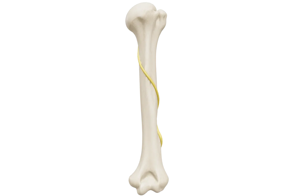
| Humeral shaft fracture, especially a spiral fracture of the distal third | Radial | Wrist and finger drop; numbness over the first dorsal web space. Most recover spontaneously |
| Shoulder dislocation or fracture of the surgical neck | Axillary | Weak abduction; numbness over the regimental badge area |
| Supracondylar fracture in a child | Anterior interosseous, radial or ulnar | Anterior interosseous injury: cannot make an ‘OK’ sign with the thumb and index finger, with no sensory loss |
| Medial epicondyle fracture or elbow trauma | Ulnar | Clawing of the ring and little fingers and weak intrinsics |
| Laceration at the wrist | Median or ulnar | Loss of thenar function or intrinsics, with sensory loss in the hand |
| Posterior hip dislocation | Sciatic (mainly the common fibular division) | Foot drop |
| Fibular neck fracture or a tight cast | Common fibular (peroneal) | Foot drop; numbness over the dorsum of the foot |
| Lymph node biopsy in the posterior triangle | Spinal accessory | Weak trapezius, shoulder droop and pain |
| Axillary surgery | Long thoracic or thoracodorsal | Winging of the scapula, or weak latissimus dorsi |
Frequently asked questions
What is the difference between neurapraxia and axonotmesis?
In neurapraxia the axon is intact and there is a local conduction block, usually from demyelination; there is no Wallerian degeneration and recovery is complete. In axonotmesis the axon is divided but its connective tissue sheath survives, so the distal axon degenerates and the nerve must regrow from the injury.
How fast do peripheral nerves regenerate?
Roughly 1 mm a day, or about an inch a month, after a delay while the axons cross the injury. The distance from injury to target therefore predicts when recovery should appear.
When does a nerve conduction study show an axonal injury?
After Wallerian degeneration is complete, at about 10 days. Before then the distal nerve still conducts and the injury can look like a neurapraxia. EMG signs of denervation appear at about two to three weeks.
What does a preserved sensory nerve action potential in an anaesthetic limb mean?
A preganglionic (root avulsion) injury. The peripheral sensory axons are still attached to their cell bodies in the dorsal root ganglion, so they conduct even though the connection to the spinal cord is lost.
When should a nerve injury be explored?
At once for a clean, sharp laceration with a deficit; at about three weeks for a contaminated or crush injury; and for a closed injury, when expected recovery fails to appear on clinical and neurophysiological review.
References
- Seddon HJ. Three types of nerve injury. Brain 1943;66(4):237–288.
- Sunderland S. A classification of peripheral nerve injuries producing loss of function. Brain 1951;74(4):491–516.
- Mackinnon SE, Dellon AL. Surgery of the Peripheral Nerve. New York: Thieme; 1988.
- Shao YC, Harwood P, Grotz MR, Limb D, Giannoudis PV. Radial nerve palsy associated with fractures of the shaft of the humerus: a systematic review. J Bone Joint Surg Br 2005;87(12):1647–1652.
- Grinsell D, Keating CP. Peripheral nerve reconstruction after injury: a review of clinical and experimental therapies. Biomed Res Int 2014;2014:698256.
Further reading
- TeachMeAnatomy — The radial nerve — Course, branches and the clinical picture of radial nerve injury
Written for revision in the MRCS Part A style under our editorial standards. It is not clinical advice: always confirm against current guidance. Spotted an error? Tell us.
Related articles
Upper limb & breast
Brachial plexus anatomy: roots to branches, and the nerve lesions that follow
Brachial plexus for MRCS Part A with a 3D model: roots, trunks, divisions, cords and branches, Erb's and Klumpke's palsies and the classic nerve lesions.
ReadUpper limb & breast
Carpal tunnel and wrist anatomy: the flexor retinaculum, nine tendons, six compartments and the snuffbox
Wrist anatomy for MRCS Part A with a 3D cross-section: flexor retinaculum, carpal tunnel contents, Guyon's canal, the six extensor compartments and snuffbox.
ReadPathology
Wound healing and inflammation: from acute inflammation to the mature scar
Acute inflammation, the phases of wound healing, collagen and tensile strength, impaired healing, keloid vs hypertrophic scar, granulomas and SSI prevention.
ReadTurn reading into marks
Drill pathology with curriculum-mapped questions, rate your confidence, and watch the gap close on your dashboard.