Brachial plexus anatomy: roots to branches, and the nerve lesions that follow
Brachial plexus for MRCS Part A with a 3D model: roots, trunks, divisions, cords and branches, Erb's and Klumpke's palsies and the classic nerve lesions.
From the neck to the axilla: how the plexus is assembled
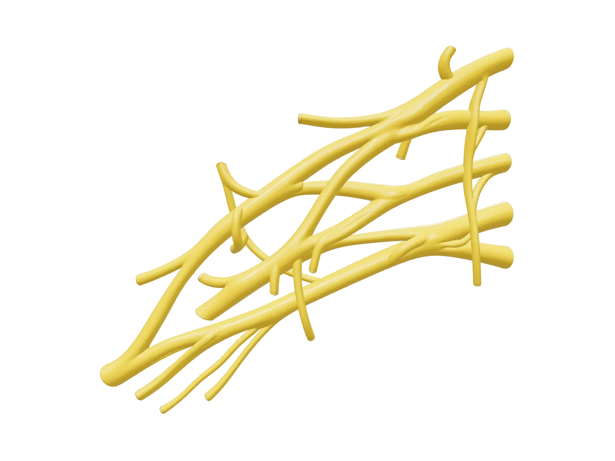
The brachial plexus is the ventral rami of C5, C6, C7, C8 and T1, which emerge between scalenus anterior and scalenus medius beneath the prevertebral fascia and reorganise four times on the way to the arm. The rule at each level is fixed. C5 and C6 unite as the upper trunk, C7 continues alone as the middle trunk and C8 and T1 unite as the lower trunk; the trunks cross the posterior triangle above the clavicle, the lower one lying behind the third part of the subclavian artery on the first rib. Behind the middle third of the clavicle each trunk splits into an anterior and a posterior division. The three posterior divisions form the posterior cord, the anterior divisions of the upper and middle trunks form the lateral cord and the anterior division of the lower trunk continues as the medial cord. The cords take their names from their position around the second part of the axillary artery, behind pectoralis minor, before giving the terminal branches. The functional logic is simpler than the diagram: anterior divisions carry fibres for the flexor compartments and posterior divisions for the extensors, so every muscle that extends the shoulder, elbow, wrist or fingers is supplied by the posterior cord through the axillary and radial nerves, and every flexor by the lateral or medial cord through the musculocutaneous, median and ulnar nerves. About half of people depart from the textbook: a prefixed plexus takes a large contribution from C4, a postfixed plexus from T2.
Interactive 3D · Brachial plexus. Tap each hotspot to name the roots, trunks, cords and branches; drag to tilt and watch the posterior divisions gather into the posterior cord.Open full screen
Branches by level with root values as given in standard texts; variation is common. The middle trunk, lower trunk and divisions give no branches.
Level
Branch
Root values
Supplies
Roots
Dorsal scapular nerve
C5
Rhomboids, levator scapulae
Roots
Long thoracic nerve
C5, C6, C7
Serratus anterior
Roots
Branch to the phrenic nerve
C5 (with C3, C4)
Diaphragm
Upper trunk
Suprascapular nerve
C5, C6
Supraspinatus, infraspinatus
Upper trunk
Nerve to subclavius
C5, C6
Subclavius
Lateral cord
Lateral pectoral nerve
C5, C6, C7
Pectoralis major (clavicular head)
Lateral cord
Musculocutaneous nerve
C5, C6, C7
Coracobrachialis (which it pierces), biceps, brachialis; lateral forearm skin
Lateral cord
Lateral root of the median nerve
C5, C6, C7
Joins the medial root in front of the axillary artery
Posterior cord
Upper subscapular nerve
C5, C6
Subscapularis
Posterior cord
Thoracodorsal nerve
C6, C7, C8
Latissimus dorsi
Posterior cord
Lower subscapular nerve
C5, C6
Subscapularis, teres major
Posterior cord
Axillary nerve
C5, C6
Deltoid, teres minor; regimental badge skin
Posterior cord
Radial nerve
C5–T1
Triceps, brachioradialis and every forearm extensor; posterior arm, forearm and radial dorsal hand skin
Medial cord
Medial pectoral nerve
C8, T1
Pectoralis minor, sternocostal pectoralis major
Medial cord
Medial cutaneous nerve of the arm
C8, T1
Medial arm skin
Medial cord
Medial cutaneous nerve of the forearm
C8, T1
Medial forearm skin
Medial cord
Ulnar nerve
C8, T1 (often C7)
Flexor carpi ulnaris, ulnar half of flexor digitorum profundus, most intrinsics; ulnar one and a half digits
Medial cord
Medial root of the median nerve
C8, T1
With the lateral root: forearm flexors and pronators, thenar muscles, lateral two lumbricals
Relations that explain the clinical picture
Roots lie between the scalenes with the phrenic nerve on the front of scalenus anterior, so an interscalene block almost always paralyses the ipsilateral hemidiaphragm and may give a transient Horner's syndrome.
Trunks cross the first rib, where the lower trunk lies behind the subclavian artery. A cervical rib or fibrous band lifts both, so thoracic outlet syndrome presents with T1 symptoms (wasting of the small hand muscles, numbness of the medial forearm) or a cold, pulseless hand.
Divisions sit behind the clavicle, protected by subclavius; they are rarely injured by a clavicular fracture, but a supraclavicular block here risks a pneumothorax.
Cords surround the second part of the axillary artery behind pectoralis minor, the medial cord between artery and vein. In an axillary node clearance the long thoracic nerve on the medial wall, the thoracodorsal bundle on the posterior wall and the intercostobrachial nerve crossing the fat are the structures at risk.
Branches each have a relation that explains their classic injury: the axillary nerve passes through the quadrangular space round the surgical neck of the humerus; the radial nerve lies in the spiral groove with the profunda brachii artery; the ulnar nerve lies behind the medial epicondyle; the median nerve runs medial to the brachial artery in the cubital fossa and then through the carpal tunnel.
Root and trunk lesions: Erb's and Klumpke's
Traction pulls the plexus in one of two directions. Forcing the head away from a depressed shoulder, in a difficult vertex delivery or a motorcyclist landing on the shoulder, tears the upper trunk at Erb's point, where C5 and C6 meet. The abductors and external rotators of the shoulder (deltoid, supraspinatus, infraspinatus), the elbow flexors (biceps, brachialis) and the supinators lose their supply, so the arm hangs adducted and internally rotated with the elbow extended and the forearm pronated: the waiter's tip. Sensation is lost over the lateral arm and forearm and the biceps and brachioradialis reflexes disappear. Pulling the abducted arm upward, when a falling person grabs a branch or a breech infant is delivered by the arm, stretches the lower roots and gives Klumpke's palsy: paralysis of the intrinsic muscles and long finger flexors produces a claw hand with wasting between the metacarpals, and sensation is lost over the medial forearm and ulnar hand. If T1 is torn from the cord proximal to the exit of its sympathetic fibres the patient also has a Horner's syndrome. A Horner's syndrome, a paralysed hemidiaphragm, a winged scapula or a preserved sensory nerve action potential in anaesthetic skin each means the injury is at root level, proximal to the dorsal root ganglion, where the roots cannot be repaired by grafting.
The two named traction patterns and the signs that place an injury at root level.
Pattern
Roots
Mechanism
Picture
Clue to the level
Erb's (upper)
C5, C6 (sometimes C7)
Head pushed away from the shoulder: shoulder dystocia, motorcycle fall
Horner's syndrome means T1 is avulsed from the cord
Whole plexus
C5–T1
High-energy traction
Flail, anaesthetic arm
Avulsions common; sensory potentials persist in numb skin
The peripheral nerve lesions Part A loves
The lesions most often set in the written paper, with the discriminating finding for each. Sensory sparing in the median and ulnar rows separates a wrist lesion from a higher one.
A 45-year-old man has anterior shoulder pain after a fall. He cannot lift the dorsum of the hand away from the lumbar spine when the arm is placed behind the back, and passive external rotation of the shoulder is increased compared with the other side.
Which structure is most likely injured?
How sure are you?
Explanation
A positive lift-off test with increased passive external rotation indicates rupture of subscapularis, the only rotator cuff muscle that inserts on the lesser tuberosity and the main medial rotator of the shoulder. Subscapularis tears often follow a fall or an anterior dislocation and may allow the long head of biceps to sublux medially, but the biceps tendon itself does not produce the lift-off sign. A supraspinatus tear weakens abduction and gives a painful arc. An infraspinatus tear weakens external rotation, producing the opposite picture of reduced active external rotation. The coracohumeral ligament reinforces the rotator interval and restrains external rotation in adduction; its injury does not abolish active medial rotation. Reference: Last's Anatomy 12e.
What are the five terminal branches of the brachial plexus and which cord gives each?+
Musculocutaneous from the lateral cord; median from the lateral and medial cords; ulnar from the medial cord; axillary and radial from the posterior cord.
How do you localise a brachial plexus injury to the roots?+
Look for the functions of branches that leave at root level: weakness of the rhomboids (dorsal scapular nerve), winging of the scapula (long thoracic nerve), a raised hemidiaphragm (phrenic contribution) or a Horner's syndrome (T1 sympathetic fibres). Any of these means the injury is proximal to the trunks.
Which nerve is injured in an anterior dislocation of the shoulder?+
The axillary nerve, as it winds round the surgical neck of the humerus through the quadrangular space. Test sensation over the regimental badge area and feel for deltoid contraction before and after reduction.
Why is the triceps spared in a radial nerve palsy from a mid-shaft humeral fracture?+
The branches to the heads of triceps leave the radial nerve in the axilla and upper arm, above the spiral groove, so a lesion in the groove paralyses the wrist and finger extensors and brachioradialis but leaves elbow extension and the triceps reflex intact.
References
Sinnatamby CS. Last's Anatomy: Regional and Applied, 12th ed. Elsevier — upper limb: the brachial plexus and its branches.
Drake RL, Vogl AW, Mitchell AWM. Gray's Anatomy for Students, 4th ed. Elsevier — upper limb: axilla and brachial plexus.
Moore KL, Dalley AF, Agur AMR. Moore's Clinically Oriented Anatomy, 9th ed. Wolters Kluwer — upper limb; clinical boxes on nerve injuries.
Williams NS, O'Connell PR, McCaskie AW (eds). Bailey & Love's Short Practice of Surgery, 28th ed. CRC Press — peripheral nerve injuries; the breast and axilla.
Written for revision in the MRCS Part A style under our editorial standards. It is not clinical advice: always confirm against current guidance. Spotted an error? Tell us.