Carpal tunnel and wrist anatomy: the flexor retinaculum, nine tendons, six compartments and the snuffbox
Wrist anatomy for MRCS Part A with a 3D cross-section: flexor retinaculum, carpal tunnel contents, Guyon's canal, the six extensor compartments and snuffbox.
The eight carpal bones lie in two rows. The proximal row, radial to ulnar, is scaphoid, lunate, triquetrum and pisiform; the distal row is trapezium, trapezoid, capitate and hamate. Together they form an arch that is concave on the palmar side, deepened at its radial edge by the tubercle of the scaphoid and the ridge of the trapezium and at its ulnar edge by the pisiform and the hook of the hamate. Across these four bony points stretches the flexor retinaculum (transverse carpal ligament), a tough band about 2.5 cm square that converts the arch into the carpal tunnel. Proximally it is continuous with the deep fascia of the forearm; distally it blends with the palmar aponeurosis, into which palmaris longus inserts across its front. The thenar muscles arise from its radial half with the scaphoid and trapezium, the hypothenar muscles from its ulnar half with the pisiform and hook of hamate. The point to hold on to is that the tunnel has bone on three sides and an unyielding ligament on the fourth, so anything that swells its contents or narrows its walls raises the pressure on the one structure inside that cannot tolerate it, the median nerve.
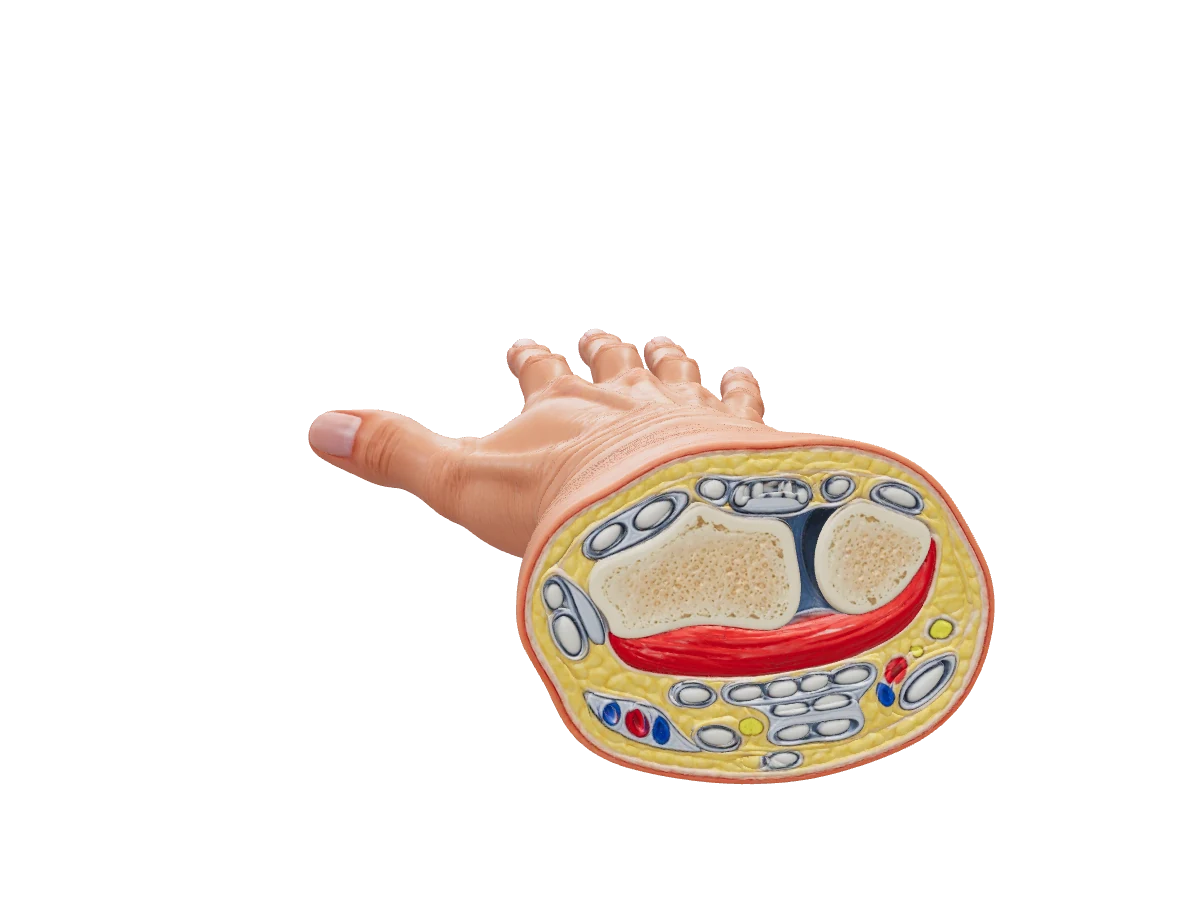
Interactive 3D · Wrist cross-section. Look into the cut face: radius and ulna with pronator quadratus in front, the flexor tendons and median nerve on the palmar side heading for the tunnel, and the six extensor compartments behind.Open full screen
What runs through the tunnel, and what runs outside it
Nine tendons. Four of flexor digitorum superficialis, four of flexor digitorum profundus and one of flexor pollicis longus. The superficialis tendons to the middle and ring fingers lie in front of those to the index and little fingers; profundus lies deepest. The finger flexors share the ulnar bursa and flexor pollicis longus has its own radial bursa; the two can communicate at the wrist, which is how infection in the thumb sheath can reach the little finger.
The median nerve enters beneath palmaris longus, in front of the superficialis tendons and slightly to their radial side, immediately under the retinaculum: the most superficial structure in the tunnel and the one the ligament presses on.
The recurrent motor branch leaves at the distal edge of the retinaculum and curves back radially to abductor pollicis brevis, opponens pollicis and the superficial head of flexor pollicis brevis. In about half of people it arises beyond the ligament, in a third beneath it and in the rest it pierces it, which is why the retinaculum is divided along its ulnar edge.
Beyond the tunnel the nerve supplies those thenar muscles and the lateral two lumbricals (LOAF) and gives sensation to the palmar skin of the thumb, index, middle and radial half of the ring finger and their nail beds.
Structures at the wrist that lie outside the carpal tunnel. Each explains a sparing sign or a surgical hazard.
Structure
Where it runs
Why it matters
Palmar cutaneous branch of the median nerve
Arises about 5 cm above the wrist crease, runs between flexor carpi radialis and palmaris longus and crosses the retinaculum superficially to the skin of the thenar eminence and central palm
Palm sensation is spared in carpal tunnel syndrome; a painful neuroma follows if a radially placed incision cuts it
Ulnar nerve and artery
Guyon's canal, between the pisiform and the hook of hamate: floor the flexor retinaculum and pisohamate ligament, roof the palmar carpal ligament and palmaris brevis. The nerve divides into a superficial sensory and a deep motor branch that hooks round the hamate
Compression paralyses the intrinsics but spares the dorsum of the hand; the artery lies radial to the nerve and continues as the superficial palmar arch
Dorsal branch of the ulnar nerve
Leaves the nerve about 5 cm above the wrist and winds beneath flexor carpi ulnaris onto the dorsum
Dorsal ulnar sensation is spared in a Guyon's canal lesion and lost in a lesion at the elbow
Flexor carpi radialis
In its own tunnel within the radial part of the retinaculum, in the groove on the trapezium
The landmark tendon: radial artery on its radial side, median nerve on its ulnar side and deeper
Palmaris longus
Superficial to the retinaculum into the palmar aponeurosis; absent in roughly one person in seven
The median nerve lies directly beneath and slightly radial to it; a useful tendon graft
Radial artery
Between flexor carpi radialis and brachioradialis, then dorsally beneath APL and EPB across the snuffbox to form the deep palmar arch; a superficial palmar branch crosses the thenar muscles
The pulse and the arterial line; Allen's test checks that the ulnar artery can supply the hand alone
Ulnar artery
Radial to the ulnar nerve, through Guyon's canal to become the main supply of the superficial palmar arch
The arch lies distal to the retinaculum, at about the level of the outstretched thumb, so decompression stops short of it
The six extensor compartments
The compartments beneath the extensor retinaculum, radial to ulnar: 2–2–1–2–1–1. Extensor indicis and extensor digiti minimi lie on the ulnar side of the communis tendon to their finger.
Radial border of the snuffbox, over the radial styloid
De Quervain's tenosynovitis, with pain on Finkelstein's test; intersection syndrome where these tendons cross the second compartment 4–6 cm above the wrist
2
Extensor carpi radialis longus and brevis
Radial to Lister's tubercle
Longus is supplied by the radial nerve above its division, so wrist extension survives a posterior interosseous palsy with radial deviation
3
Extensor pollicis longus
Hooks round the ulnar side of Lister's tubercle; ulnar border of the snuffbox
Attrition rupture a few weeks after an undisplaced distal radius fracture: the thumb can no longer lift off a flat table
4
Extensor digitorum, extensor indicis
In line with the third metacarpal
The terminal branch of the posterior interosseous nerve lies on the floor and supplies the wrist joint; the usual dorsal approach to the wrist
5
Extensor digiti minimi
Over the distal radio-ulnar joint
Ruptures in rheumatoid synovitis of the joint (Vaughan-Jackson), the little finger dropping first
6 (most ulnar)
Extensor carpi ulnaris
In the groove on the back of the distal ulna, held by its own subsheath
Part of the triangular fibrocartilage complex; subluxes out of its groove in forced supination
The extensor retinaculum runs obliquely from the radius to the pisiform and triquetrum rather than to the ulna, so the forearm can rotate beneath it, and sends septa to the bone that make the six tunnels, each with its own synovial sheath. The superficial branch of the radial nerve crosses the first compartment in the subcutaneous tissue here, which is why tight watch straps and handcuffs numb the back of the thumb web.
The anatomical snuffbox and the scaphoid
Radial (anterior) border
The tendons of abductor pollicis longus and extensor pollicis brevis, the first extensor compartment.
Ulnar (posterior) border
The tendon of extensor pollicis longus, having turned round Lister's tubercle.
Floor
The radial styloid proximally, then the scaphoid and trapezium, then the base of the first metacarpal. The scaphoid is the bone you press on.
Contents
The radial artery on the floor, crossing from palmar to dorsal; the cephalic vein and the terminal branches of the superficial radial nerve in the roof.
The scaphoid is the commonest carpal bone to fracture, usually across its waist after a fall on the outstretched hand, and its blood supply explains why the fracture matters. Branches of the radial artery enter through the dorsal ridge near the distal pole and run proximally inside the bone, so the proximal pole depends on flow across the waist; a displaced waist or proximal pole fracture cuts it off, with avascular necrosis and non-union to follow. The signs are tenderness in the snuffbox and over the scaphoid tubercle and pain on axial compression of the thumb, and the first radiographs are often normal. NICE guidance on non-complex fractures advises considering MRI as the first-line investigation when a scaphoid fracture is suspected and the films do not show it. One other injury belongs here: the lunate, in the floor of the tunnel, can dislocate forward into it after a hyperextension injury and present as an acute carpal tunnel syndrome.
Carpal tunnel syndrome and the anatomy of decompression
Carpal tunnel syndrome is the commonest entrapment neuropathy. The patient, typically a middle-aged woman, is woken by tingling and pain in the thumb, index and middle fingers and shakes the hand to relieve it; holding a phone or a steering wheel provokes it and the ache may spread up the forearm. Later the thumb becomes clumsy, the thenar eminence flattens as abductor pollicis brevis wastes, and objects are dropped. The sensory loss stops at the wrist crease: the palm, supplied by the palmar cutaneous branch, is normal, which separates the tunnel from a lesion in the forearm. Tapping the nerve at the crease sends tingling into the fingers (Tinel's), a minute of full wrist flexion reproduces the symptoms (Phalen's) and so does pressure over the tunnel (Durkan's); nerve conduction studies confirm a prolonged latency across the wrist. The causes are anything that fills the tunnel or narrows it: pregnancy, hypothyroidism, rheumatoid tenosynovitis, diabetes, acromegaly, dialysis amyloid, obesity, a malunited distal radius fracture, a ganglion. Night splinting and a steroid injection help many patients; the definitive treatment is division of the flexor retinaculum, open or endoscopic.
Find the landmarks. The distal wrist crease marks the proximal edge of the retinaculum. The pisiform is the knob at the ulnar end of the crease, the hook of hamate about 2 cm distal and radial to it; the scaphoid tubercle is at the radial end, the ridge of the trapezium just beyond.
Kaplan's cardinal line, from the apex of the thumb web parallel to the proximal palmar crease towards the hook of hamate, marks the distal edge of the retinaculum, the level of the superficial palmar arch and the emergence of the recurrent motor branch. The release does not go beyond it.
The incision runs from just distal to the wrist crease along a line continued from the radial border of the ring finger, keeping ulnar to the thenar crease: ulnar to the palmar cutaneous and recurrent motor branches, radial to the ulnar neurovascular bundle in Guyon's canal.
Divide the retinaculum under direct vision along its ulnar side, from the palmar aponeurosis back to the forearm fascia, until the nerve is free along its whole length and the fat of the palm bulges into the wound.
The complications map onto the anatomy: incomplete release proximally, a neuroma of the palmar cutaneous branch, weak thumb abduction from damage to the motor branch, bleeding from the superficial arch distally, and pillar pain at the thenar and hypothenar origins.
A 45-year-old man has anterior shoulder pain after a fall. He cannot lift the dorsum of the hand away from the lumbar spine when the arm is placed behind the back, and passive external rotation of the shoulder is increased compared with the other side.
Which structure is most likely injured?
How sure are you?
Explanation
A positive lift-off test with increased passive external rotation indicates rupture of subscapularis, the only rotator cuff muscle that inserts on the lesser tuberosity and the main medial rotator of the shoulder. Subscapularis tears often follow a fall or an anterior dislocation and may allow the long head of biceps to sublux medially, but the biceps tendon itself does not produce the lift-off sign. A supraspinatus tear weakens abduction and gives a painful arc. An infraspinatus tear weakens external rotation, producing the opposite picture of reduced active external rotation. The coracohumeral ligament reinforces the rotator interval and restrains external rotation in adduction; its injury does not abolish active medial rotation. Reference: Last's Anatomy 12e.
What are the attachments of the flexor retinaculum?+
On the ulnar side, the pisiform and the hook of hamate; on the radial side, the tubercle of the scaphoid and the ridge of the trapezium. It bridges the carpal arch to form the carpal tunnel and gives origin to the thenar and hypothenar muscles.
What passes through the carpal tunnel?+
Nine tendons and the median nerve: four of flexor digitorum superficialis, four of flexor digitorum profundus and flexor pollicis longus. The ulnar nerve and artery, palmaris longus, flexor carpi radialis (in its own tunnel) and the palmar cutaneous branch of the median nerve all pass outside it.
What are the six extensor compartments of the wrist?+
From radial to ulnar: 1, abductor pollicis longus and extensor pollicis brevis; 2, extensor carpi radialis longus and brevis; 3, extensor pollicis longus; 4, extensor digitorum and extensor indicis; 5, extensor digiti minimi; 6, extensor carpi ulnaris.
Why is palm sensation preserved in carpal tunnel syndrome?+
Because the palmar cutaneous branch of the median nerve leaves the main trunk about 5 cm above the wrist and crosses the flexor retinaculum superficially, outside the tunnel, so it is not compressed. Numbness over the palm points to a lesion higher in the forearm.
References
Sinnatamby CS. Last's Anatomy: Regional and Applied, 12th ed. Elsevier — upper limb: the wrist, carpal tunnel and hand.
Drake RL, Vogl AW, Mitchell AWM. Gray's Anatomy for Students, 4th ed. Elsevier — upper limb: wrist and hand, carpal tunnel and extensor compartments.
Moore KL, Dalley AF, Agur AMR. Moore's Clinically Oriented Anatomy, 9th ed. Wolters Kluwer — upper limb; clinical boxes on carpal tunnel syndrome and scaphoid fracture.
Williams NS, O'Connell PR, McCaskie AW (eds). Bailey & Love's Short Practice of Surgery, 28th ed. CRC Press — the hand; upper limb injuries.
Written for revision in the MRCS Part A style under our editorial standards. It is not clinical advice: always confirm against current guidance. Spotted an error? Tell us.